Mastering Pterygoid Implant Placement: An Anatomical Guide
January 16, 2026
Pterygoid Implant Anatomy: What Patients Should Know
Pterygoid implants are a specialized dental implant option used in the posterior upper jaw. Their placement is different from that of conventional implants because they are designed to obtain support from dense bone located farther back in the maxilla.
Understanding pterygoid implant anatomy helps explain why this treatment may be considered for patients who have limited bone in the posterior maxilla and why detailed three-dimensional planning is important before treatment.
Understanding the Anatomy Behind Pterygoid Implants
The posterior upper jaw can present challenges for implant placement. After teeth are lost, the amount and shape of available bone may change over time. The nearby maxillary sinus can also limit the vertical bone available for conventional implants.
Pterygoid implants use a different anatomical pathway. Rather than depending entirely on the bone directly beneath the missing teeth, they extend toward dense bone in the posterior maxillary region.
Key anatomical structures include:
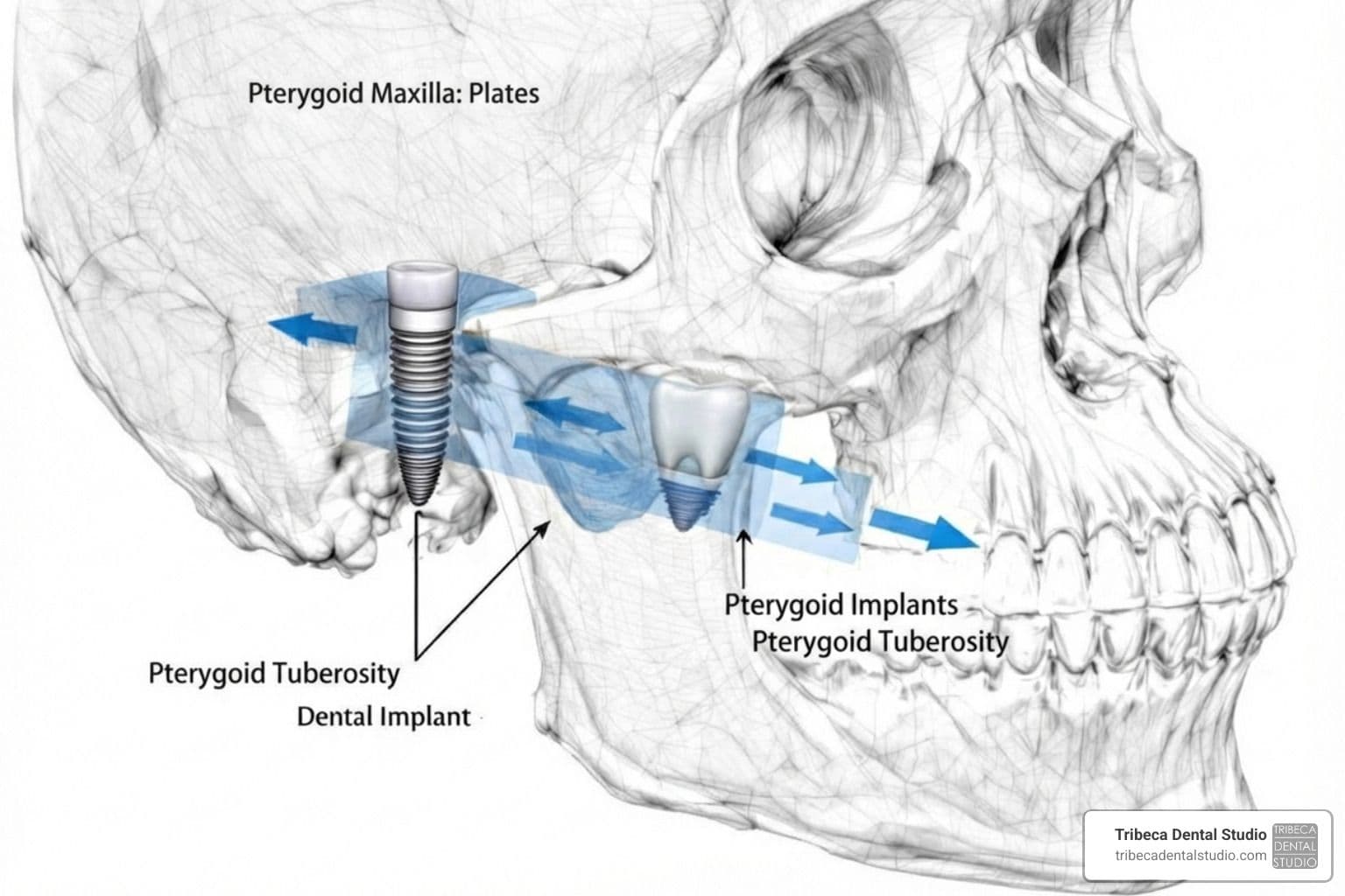
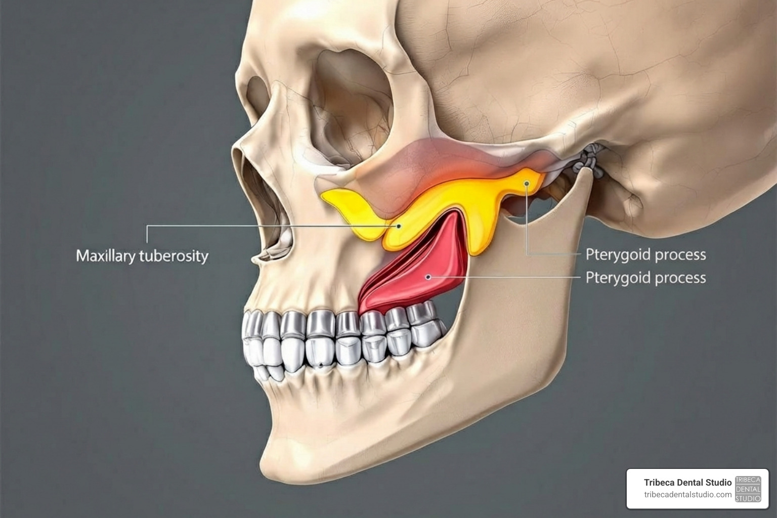
- Maxillary Tuberosity – the rounded posterior portion of the upper jaw and a common starting region for the implant trajectory.
- Pyramidal Process of the Palatine Bone – an intermediate bony structure located behind the maxillary tuberosity.
- Pterygoid Process of the Sphenoid Bone – a dense posterior structure that can contribute to implant anchorage.
- Greater Palatine Canal – an important nearby anatomical structure containing nerves and blood vessels that must be considered during treatment planning.
Because these structures are located close together, pterygoid implant treatment requires careful evaluation of each patient's individual anatomy.
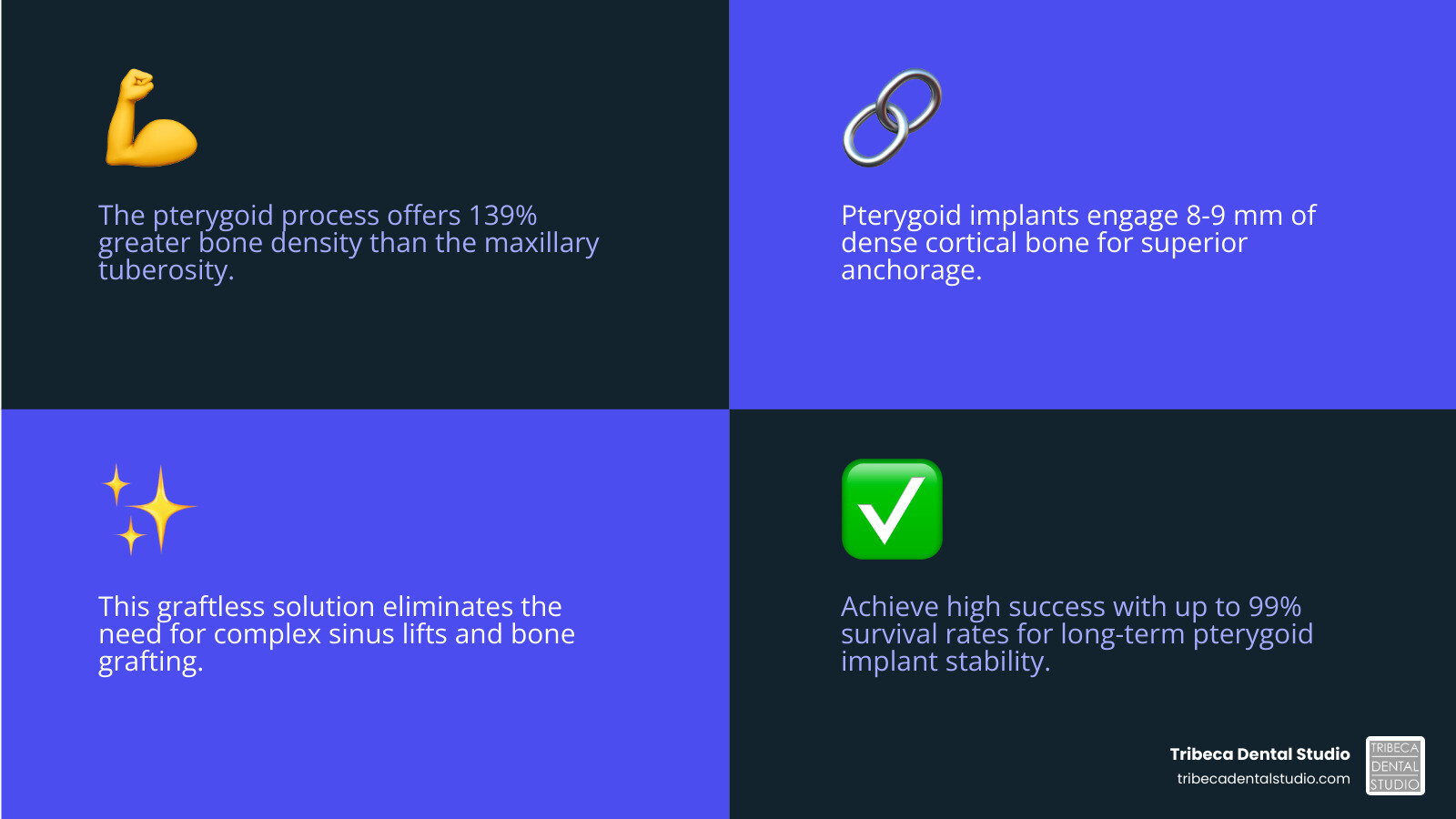
The Core of Pterygoid Implant Anatomy
The pterygomaxillary region is located toward the back of the upper jaw. It includes several closely related bones and anatomical spaces that may influence implant planning.
A pterygoid implant may pass through or engage portions of the maxillary tuberosity and palatine bone before obtaining support in the pterygoid region. The exact trajectory varies according to the patient's anatomy, available bone, restorative plan, and clinician's treatment approach.
For additional anatomical background, see this anatomical study of pterygoid implants, arteries, and nerves .
Patients who want to understand other approaches for rebuilding insufficient bone can also review our Bone Grafting services .
Key Anatomical Structures for Placement
Several bony landmarks are particularly relevant when evaluating the posterior upper jaw for pterygoid implants.
- Maxillary Tuberosity: The rounded posterior area of the maxilla. Its size, shape, and bone quality vary from person to person.
- Pyramidal Process of the Palatine Bone: Located posterior to the maxillary tuberosity, this structure may contribute to the bony pathway used during implant placement.
- Pterygoid Process of the Sphenoid Bone: Located deeper in the posterior facial skeleton, this region contains relatively dense cortical bone that may provide support for appropriately planned implants.
- Pterygoid Fossa: A depression located between portions of the pterygoid plates. Understanding its relationship to surrounding structures helps clinicians orient the implant pathway.
The goal of treatment planning is not simply to place a long implant. The implant must follow an appropriate three-dimensional path while considering bone availability, nearby anatomical structures, restorative requirements, and long-term access for hygiene.
Understanding Bone Density and Morphology
Bone quality can vary considerably throughout the upper jaw. The posterior maxilla may contain relatively soft bone, particularly in areas where teeth have been missing for a long period.
The deeper pterygoid region contains cortical bone that may provide additional support when conventional posterior implant placement is difficult. This anatomical difference is one reason pterygoid implants may be considered in selected cases.
However, there is significant variation between patients. Bone dimensions, sinus anatomy, degree of bone loss, tooth-loss history, age, and other factors can affect treatment planning.
For this reason, published anatomical averages should not be used as a substitute for patient-specific imaging and clinical evaluation.
Navigating the Greater Palatine Canal
One of the important structures near the pterygoid implant pathway is the Greater Palatine Canal.
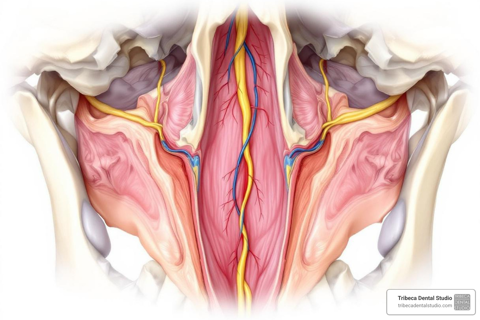
The canal carries neurovascular structures, including branches associated with the descending palatine artery and greater palatine nerve. Because of its location in the posterior maxilla, it is important to identify its position during treatment planning.
The anatomy of this region can vary significantly. Three-dimensional imaging can help the clinician evaluate the relationship between the proposed implant pathway, surrounding bone, the maxillary sinus, and nearby neurovascular structures.
Rather than relying on a fixed anatomical measurement for every patient, implant planning should be based on the patient's own imaging and clinical findings.
Angulation and Imaging for Pterygoid Implants
Pterygoid implants differ from conventional dental implants partly because their trajectory is angled toward the posterior anatomical structures of the upper jaw.
There is no single implant angle that is appropriate for every patient. Published anatomical and clinical studies describe different trajectories based on the relationship between the maxilla, palatine bone, pterygoid region, occlusal plane, and other anatomical reference points.
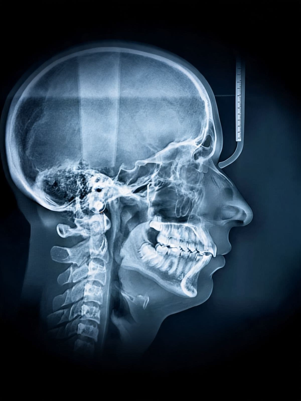
For this reason, modern planning commonly relies on Cone Beam Computed Tomography (CBCT).
CBCT imaging provides a three-dimensional view of:
- available bone volume,
- bone shape and orientation,
- the maxillary sinus,
- the posterior maxillary region,
- nearby neurovascular structures, and
- the proposed restorative position.
These images can help clinicians evaluate whether a pterygoid implant is anatomically appropriate and determine a potential implant length, position, and trajectory based on the individual case.
For more information about three-dimensional treatment planning, visit our page on Guided Dental Implant Surgery .
Potential Advantages of Pterygoid Implants
Pterygoid implants may be considered when there is limited bone in the posterior maxilla and conventional implants would otherwise require additional procedures.
A Potential Alternative to Sinus Augmentation
The maxillary sinus can limit available bone height in the posterior upper jaw. In conventional implant treatment, some patients may require a sinus augmentation procedure before or during implant placement.
Because a pterygoid implant follows a different anatomical pathway, it may allow clinicians to avoid or reduce the need for sinus augmentation in selected cases.
Whether this is appropriate depends on the patient's bone anatomy and overall treatment plan.
Patients can learn more about conventional augmentation on our Sinus Lift services page.
Potential to Reduce Bone Grafting in Selected Cases
Pterygoid implants are designed to obtain anchorage from existing posterior bone. In some cases, this may reduce the need for extensive grafting before implant placement.
However, not every patient with posterior bone loss is a candidate for this technique. Some patients may still benefit from bone grafting, sinus augmentation, alternative implant designs, or a different restorative approach.
Posterior Support for Full-Arch Restorations
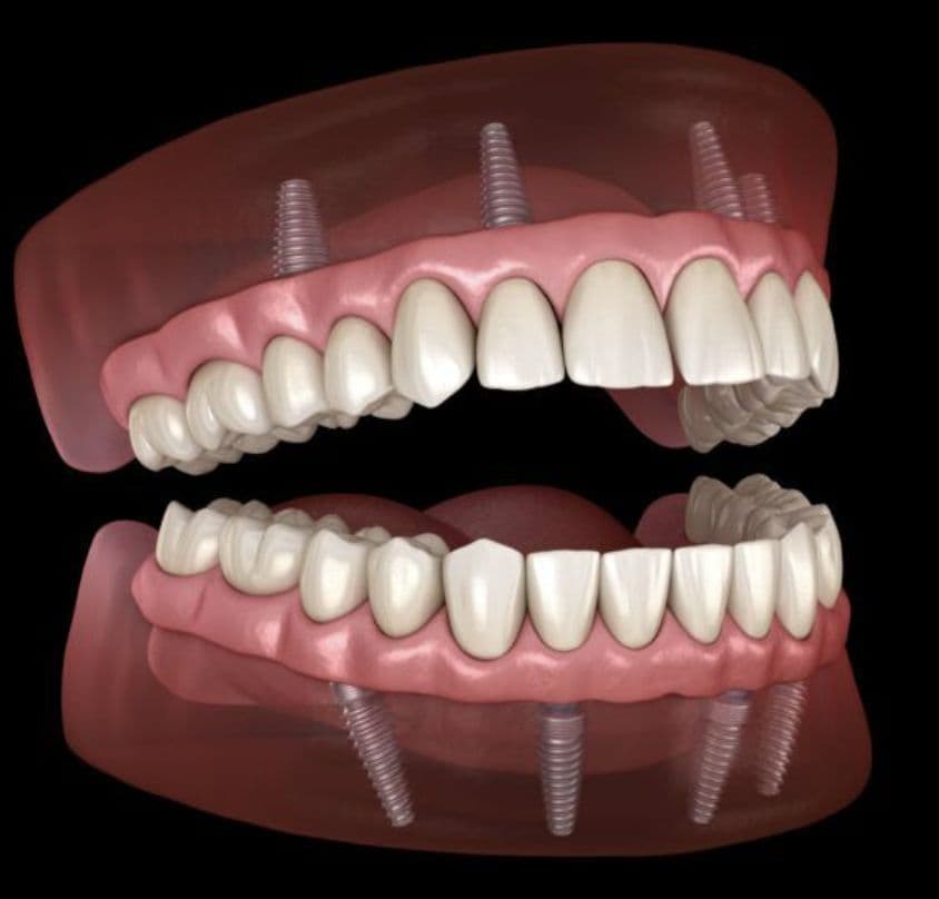
One potential restorative advantage of placing implants farther back in the upper jaw is improved posterior support for a fixed prosthesis.
In selected full-arch treatment plans, posterior implant support may help reduce the length of a distal cantilever and distribute forces across the restoration.
The final restorative design depends on the number and position of implants, bone anatomy, bite forces, prosthetic space, and other clinical considerations.
The American College of Prosthodontists has published additional information regarding implants placed in the pterygoid region: Position Statement: Use of Implants in the Pterygoid Region for Prosthodontic Treatment .
Clinical Outcomes and Survival
Clinical studies and systematic reviews have reported favorable survival outcomes for pterygoid implants in appropriately selected patients.
However, published results vary according to implant design, surgical technique, follow-up period, patient population, prosthetic design, and study methodology.
For patients, the more useful question is not whether a particular percentage applies universally, but whether this treatment is appropriate for their individual anatomy and restorative needs.
Long-term implant outcomes may also depend on oral hygiene, periodontal health, smoking status, bite forces, maintenance, medical history, prosthetic design, and regular follow-up care.
Risks, Complications, and Patient Selection
Pterygoid implant placement is an advanced procedure that requires detailed anatomical assessment and careful treatment planning.
Potential considerations include:
- limited surgical access in the posterior mouth,
- variation in posterior maxillary anatomy,
- proximity to blood vessels and nerves,
- the need for accurate implant trajectory,
- prosthetic positioning,
- difficulty accessing very posterior areas for hygiene, and
- the need for long-term implant and prosthetic maintenance.
As with other implant procedures, risks may include infection, bleeding, discomfort, failure of osseointegration, damage to nearby structures, prosthetic complications, and the possibility that additional treatment may be needed.
The presence and magnitude of these risks depend on the individual clinical situation.
When Might Pterygoid Implants Be Considered?
Pterygoid implants may be considered as part of a broader implant treatment plan when conventional posterior implant placement is limited by bone availability.
| Situations Where They May Be Considered | Situations Requiring Additional Evaluation |
|---|---|
| Reduced Posterior Maxillary Bone: Limited bone in the molar region may make conventional implants difficult without augmentation. | Limited Mouth Opening: Restricted access may make treatment technically difficult. |
| Posterior Tooth Loss: Additional posterior support may be useful in selected restorative plans. | Unfavorable Posterior Anatomy: Individual anatomical variations may make the proposed pathway unsuitable. |
| Full-Arch Rehabilitation: Pterygoid implants may be incorporated into some full-arch implant designs. | Active Infection: Infection in the proposed treatment area generally requires evaluation and management before implant placement. |
| Desire to Evaluate Alternatives to Grafting: Some patients may want to explore treatment strategies that use existing posterior bone. | Medical Conditions Affecting Surgery or Healing: Medical history and medications must be reviewed before implant treatment. |
| Need for More Posterior Implant Support: Posterior anchorage may be useful for certain prosthetic designs. | Anatomical Obstructions: Impacted teeth, unusual bone anatomy, or nearby structures may affect treatment feasibility. |
The decision to use pterygoid implants should therefore be based on clinical examination, diagnostic imaging, medical history, restorative objectives, and discussion of alternative treatments.
Pterygoid vs. Zygomatic Implants
Pterygoid and zygomatic implants are both advanced implant approaches that may be considered when there is significant bone loss in the upper jaw, but they use different anatomical regions.
Pterygoid Implants
Pterygoid implants obtain support from structures in the posterior maxillary and pterygoid region.
They may be considered when posterior maxillary bone is limited but an appropriate pathway toward the pterygoid region remains available.
Zygomatic Implants
Zygomatic implants are considerably longer implants that obtain anchorage from the zygomatic bone, or cheekbone.
They are commonly discussed in the context of advanced maxillary bone loss when conventional implant placement may be difficult or impossible without extensive reconstruction.
How the Approaches Differ
The two treatments differ in implant length, anatomical pathway, surgical access, restorative planning, and the structures involved.
Neither option is universally preferable. The appropriate treatment depends on individual anatomy, degree and location of bone loss, restorative needs, medical history, and other factors.
For additional information, visit our page about Zygomatic Dental Implants .
Frequently Asked Questions About Pterygoid Implants
Why might pterygoid implants be considered as an alternative to a sinus lift?
Conventional implants in the posterior upper jaw require sufficient bone beneath the maxillary sinus.
When that bone is limited, sinus augmentation may be one treatment option. Pterygoid implants follow a different pathway toward posterior bone and therefore may provide another option in selected cases.
The choice between these approaches depends on individual anatomy and the overall restorative plan.
How successful are pterygoid implants?
Published studies generally report favorable survival outcomes for pterygoid implants, but the exact results vary between studies and patient populations.
No published success percentage can predict the outcome for an individual patient.
Factors such as bone anatomy, implant position, oral hygiene, general health, smoking, bite forces, prosthetic design, maintenance, and follow-up can influence long-term outcomes.
What is most important about pterygoid implant anatomy?
One of the most important considerations is the three-dimensional relationship between the proposed implant pathway and the surrounding bone, maxillary sinus, greater palatine canal, and other nearby structures.
Because anatomy varies between patients, CBCT imaging can be valuable for evaluating the posterior maxilla and planning an implant trajectory based on the individual's anatomy rather than relying only on general anatomical averages.
Do pterygoid implants always eliminate the need for bone grafting?
No. Pterygoid implants may reduce or avoid the need for certain augmentation procedures in selected cases, but they do not eliminate bone grafting for every patient.
The amount and location of bone loss, implant strategy, restorative goals, and the anatomy of the posterior maxilla all influence the treatment plan.
Are pterygoid implants appropriate for everyone?
No. These implants are used for specific clinical situations.
A comprehensive evaluation is needed to determine whether conventional implants, bone grafting, sinus augmentation, pterygoid implants, zygomatic implants, or another restorative option is appropriate.
Understanding Your Options for Posterior Upper-Jaw Implants
Pterygoid implants provide an alternative anatomical approach to dental implant placement in the posterior upper jaw.
Instead of relying only on bone directly below the maxillary sinus, the implant is planned toward posterior structures that may provide additional support in appropriately selected patients.
The key to understanding this treatment is anatomy. The maxillary tuberosity, palatine bone, pterygoid region, maxillary sinus, and nearby neurovascular structures must all be considered when evaluating a potential implant pathway.
CBCT imaging and comprehensive restorative planning can help determine whether this approach is appropriate for a particular case.
Patients with significant posterior upper-jaw bone loss may have several treatment options, including conventional implants with augmentation, pterygoid implants, zygomatic implants, or other restorative approaches.
To learn more about advanced implant treatment options, visit our Zygomatic Dental Implants page or speak with Tribeca Dental Studio about which options may be appropriate for your individual situation.